
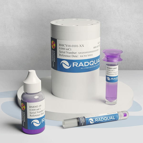
PET F-18 X-Cal System
This patented, RadQual exclusive product allows for cross calibration of your PET scanner, dose calibrator, and well detector for Ga-68 and F-18, and is useful in multi-center imaging trials to both assess bias and enable correction of biases due to instrumentation factors for serial PET studies.
- Cylinder contains a 0.5 mCi (18.5 MBq) of Ge-68/Ga-68 (in secular equilibrium) which is implicitly traceable to NIST and is supplied with a base mount for the Data Spectrum ECT Phantom.
- The dose calibrator for this series contains approximately 25 µCi (0.90 MBq) of F-18 equivalent activity and is directly traceable to NIST.
- The rod source contains approximately 0.14 µCi (3.85 kBq) and is implicitly traceable to NIST. A custom decay chart for Ge-68/Ga-68 and F-18 are provided with this source series.
Why Radqual Pet F-18 X-Cal System?
All of the sources in this set are manufactured from the same Ge68/Ga-68 epoxy process using NIST traceable balances to ensure accurate measurement of weight. Our proprietary process allows for extremely uniform activity distribution within the cylinder. The content of the cylinder and rod source are determined by the concentration of the directly traceable dose calibrator standard. So the activity contents of all of the sources are known at the 95% confidence level within +/- 2.5%.
Research & Publications
Doot, R. K., Thompson, T., Greer, B. E., Allberg, K. C., Linden, H. M., Mankoff, D. A., & Kinahan, P. E. (2012). Early experiences in establishing a regional quantitative imaging network for PET/CT clinical trials. Magnetic resonance imaging, 30(9), 1291–1300.
https://www.sciencedirect.com/science/article/abs/pii/S0730725X12002007?via%3Dihub
Byrd, D., Christopfel, R., Arabasz, G., Catana, C., Karp, J., Lodge, M. A., Laymon, C., Moros, E. G., Budzevich, M., Nehmeh, S., Scheuermann, J., Sunderland, J., Zhang, J., & Kinahan, P. (2018). Measuring temporal stability of positron emission tomography standardized uptake value bias using long-lived sources in a multicenter network. Journal of medical imaging (Bellingham, Wash.), 5(1), 011016.
https://www.ncbi.nlm.nih.gov/pmc/articles/PMC5753626/
Doot, R. K., Pierce, L. A., 2nd, Byrd, D., Elston, B., Allberg, K. C., & Kinahan, P. E. (2014). Biases in Multicenter Longitudinal PET Standardized Uptake Value Measurements. Translational oncology, 7(1), 48–54.
https://www.ncbi.nlm.nih.gov/pmc/articles/PMC3998681/
Byrd, D. W., Sunderland, J. J., Lee, T. C., & Kinahan, P. E. (2019). Bias in PET Images of Solid Phantoms Due to CT-Based Attenuation Correction. Tomography (Ann Arbor, Mich.), 5(1), 154–160.
https://www.ncbi.nlm.nih.gov/pmc/articles/PMC6403023/
Byrd, D. W., Doot, R. K., Allberg, K. C., MacDonald, L. R., McDougald, W. A., Elston, B. F., Linden, H. M., & Kinahan, P. E. (2016). Evaluation of Cross-Calibrated 68Ge/68Ga Phantoms for Assessing PET/CT Measurement Bias in Oncology Imaging for Single- and Multicenter Trials. Tomography (Ann Arbor, Mich.), 2(4), 353–360.
https://www.ncbi.nlm.nih.gov/pmc/articles/PMC5214172/
MacDonald, L. R., Perkins, A. E., & Tung, C. H. (2017). Longitudinal monitoring of reconstructed activity concentration on a clinical time-of-flight PET/CT scanner. Journal of medical imaging (Bellingham, Wash.), 4(1), 011004.
https://www.ncbi.nlm.nih.gov/pmc/articles/PMC5120216/
Lodge, M. A., Holt, D. P., Kinahan, P. E., Wong, D. F., & Wahl, R. L. (2015). Performance assessment of a NaI(Tl) gamma counter for PET applications with methods for improved quantitative accuracy and greater standardization. EJNMMI physics, 2, 11.
https://www.ncbi.nlm.nih.gov/pmc/articles/PMC4452125/
Press, R. H., Shu, H. G., Shim, H., Mountz, J. M., Kurland, B. F., Wahl, R. L., Jones, E. F., Hylton, N. M., Gerstner, E. R., Nordstrom, R. J., Henderson, L., Kurdziel, K. A., Vikram, B., Jacobs, M. A., Holdhoff, M., Taylor, E., Jaffray, D. A., Schwartz, L. H., Mankoff, D. A., Kinahan, P. E., … Buatti, J. M. (2018). The Use of Quantitative Imaging in Radiation Oncology: A Quantitative Imaging Network (QIN) Perspective. International journal of radiation oncology, biology, physics, 102(4), 1219–1235.
https://www.ncbi.nlm.nih.gov/pmc/articles/PMC6348006/
Mira, K., Wagatsuma, K., Iimori, T., Sawada, K., Kamiya, T., Samurai, M., Miyaji, N., Murata, T., & Sato, E. (2018). Multicenter study of quantitative PET system harmonization using NIST-traceable 68Ge/68Ga cross-calibration kit. Physica Medica, 52, 98-103.
https://www.sciencedirect.com/science/article/abs/pii/S1120179718311256
| PET F-18 X-Cal System Specifications | ||
|---|---|---|
| Cylinder | Overall Dimensions | 9.37 cm (3.68 inches) height 7.04 cm (2.77 inches) diameter |
| Active Matrix | 4.50 cm (1.77 inches) height 4.50 cm (1.77 inches) diameter |
|
| Base Mount | Overall Dimensions | 18.0 cm (7.08 inches) diameter |